A Stress Hormone May Help the Brain Repair Itself
A Groundbreaking Discovery in Brain Repair
When laboratory mice experience brain damage, researchers at the Max Planck Institute of Psychiatry have repeatedly observed a fascinating response. A particular group of cells appears and becomes active around the damaged area. Jan Deussing, a research group leader and experienced neurobiologist, had observed this phenomenon many times, but he did not know exactly what type of cells were involved.
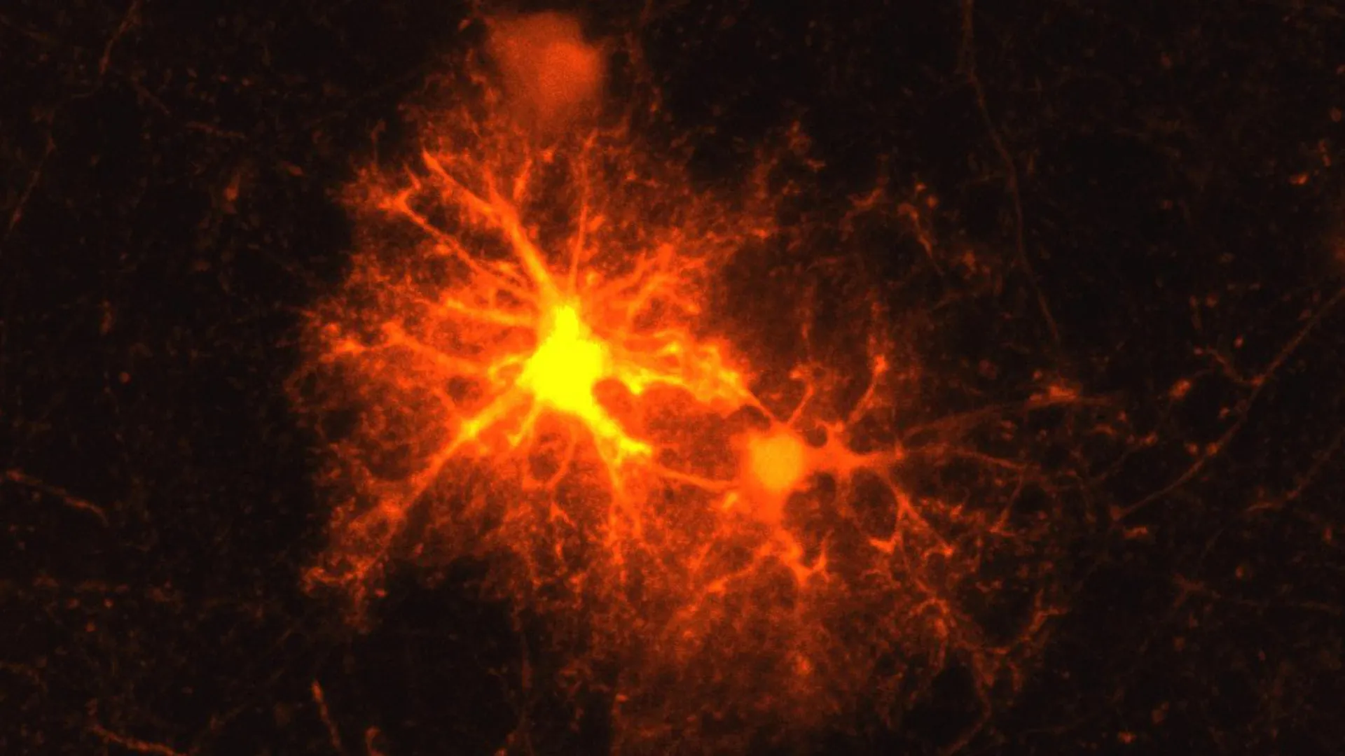
The mystery became an ideal research question for a master’s student. Clemens Ries, who had recently joined the Max Planck Institute of Psychiatry for an internship, took on the challenge. Using a mouse model, Ries systematically tested markers for all known cell types. Only one produced a response: the marker for oligodendrocyte progenitor cells (OPCs).
These precursor cells can mature into oligodendrocytes, which produce the myelin sheath surrounding axons. Axons are extensions of nerve cells that allow neurons to communicate with one another. Myelin acts much like the insulating material around an electrical cable, supporting efficient information transmission along axons and helping supply them with nutrients.
Damage to myelin can have serious consequences. In autoimmune diseases such as multiple sclerosis (MS), the protective coating breaks down. Physical injuries can also harm myelin, and in severe cases, the resulting damage can lead to the death of entire neurons. Restoring myelin around affected axons is therefore an important part of the brain’s response to injury.
A Surprising Stress Hormone Appears After Injury
Ries initially studied the newly identified cells for his master’s thesis.